
Protein Viewer plugin
Short answer: Open the Protein tab, type a PDB ID such as 1HHO and press Go. It is installed by default and works without signing in.
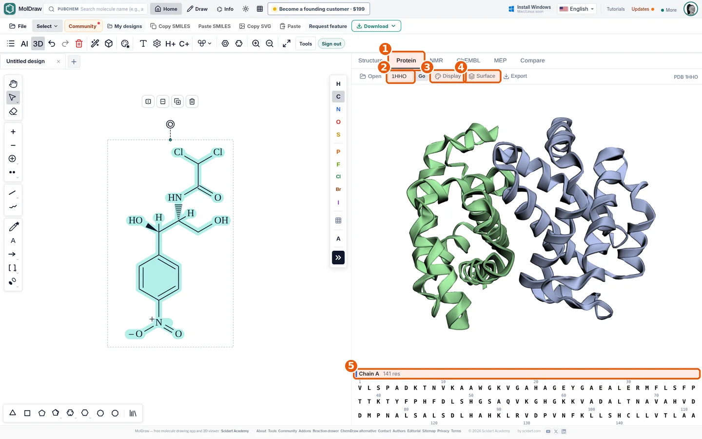
- Protein tab.
- PDB ID box: type an ID and press Go (or use Open for a file).
- Display menu: cartoon, sticks, lines, spheres and colouring.
- Surface: molecular surface with opacity and colour.
- Sequence strip: click residues to select and style them.
What it does#
Open the Protein tab next to Structure. Type a PDB ID like 1HHO, or load a file.
- Display sets the style: cartoon, stick or surface.
- Color by chain, secondary structure or element.
- Click the sequence strip to select residues.
- Export a PNG of the view.
Who it is for#
Biochemistry students. Teachers who need protein figures. Researchers who want a quick look at a structure.
How to use it#
- Open the editor (no sign-in needed) and click the Protein tab (callout 1).
- Type a PDB ID, e.g.
1HHO, and press Go (callout 2), or press Open to load a local file. - Use Display (callout 3) to switch cartoon/sticks and colour by chain.
- Add a Surface (callout 4) and lower its opacity to see the backbone.
- Click residues in the sequence strip (callout 5) to highlight them.
- Press Export to download an image.
Tips#
- Try 1HHO (haemoglobin), 1BNA (B-DNA), 6LU7 (SARS-CoV-2 main protease) or 1GFL (GFP).
- Calculate a protein's mass from its sequence with the protein molecular weight calculator.
- For docking prep, use PDB to PDBQT.
Questions and answers
Do I need an account to view proteins?
No. The Protein Viewer is installed by default and works without signing in.
Which file formats can I open?
PDB IDs are fetched directly; local PDB and mmCIF files can be opened from your computer.
Can I show a ligand as sticks?
Yes. Select the residue or ligand in the sequence strip and change its style.