View Protein Structures from the PDB in 3D with the MolDraw Protein Viewer
Short answer: Open the Protein tab next to Structure, type a four-character PDB ID such as 1HHO and press Go. The structure loads as a cartoon coloured by chain. Use Display and Surface to change the view, select residues in the sequence strip to style them, and use Export to save an image or a file. The viewer is on by default and needs no account.
Why look at a protein in 3D?
A protein's function depends on its shape. The amino-acid sequence folds into helices and sheets, and these pack into a three-dimensional structure with grooves and pockets. A small molecule drug usually works by fitting into one of those pockets. You cannot see any of that in a sequence or a 2D drawing.
The Protein Data Bank (PDB) is the public archive of experimentally determined structures of proteins, DNA and RNA. Each entry has a four-character ID, such as 1HHO for haemoglobin. The MolDraw Protein viewer loads these entries next to your drawing, so you can move from a ligand to its target without opening another program.
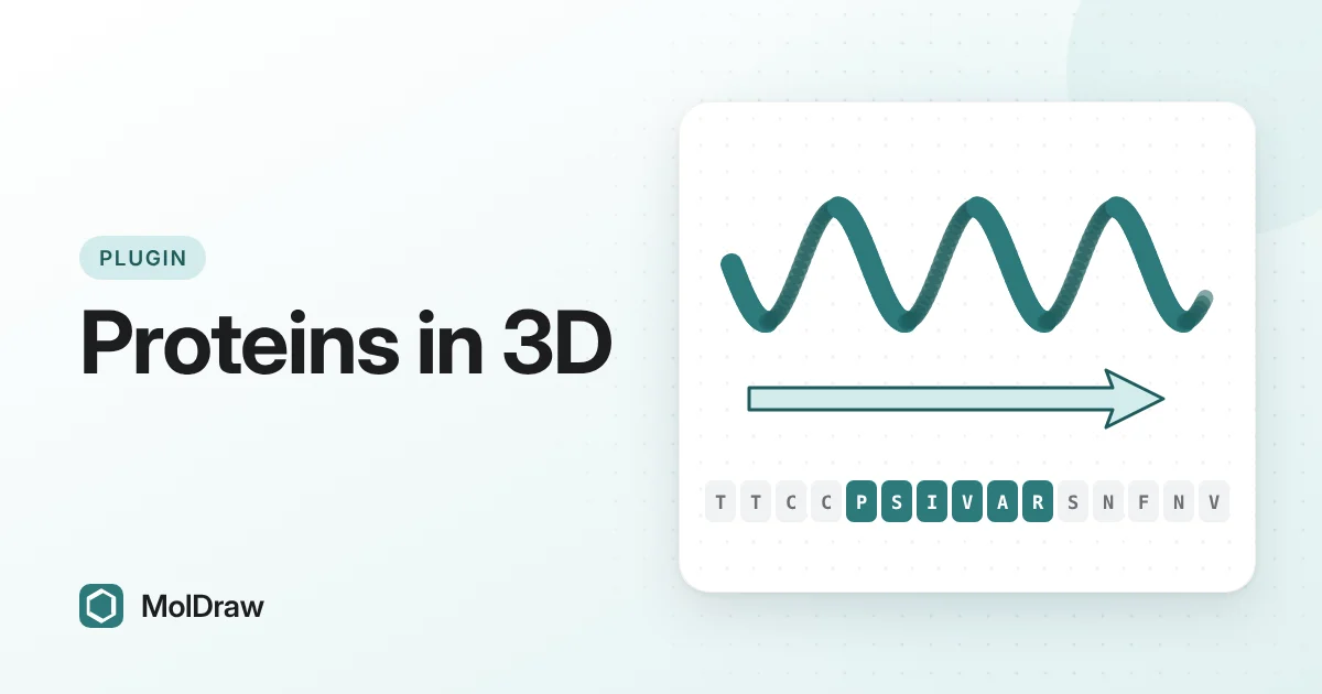
Different views answer different questions. A cartoon traces the backbone and shows secondary structure: helices as spirals and β-strands as arrows. Sticks show individual atoms, which you need for side chains and binding contacts. A surface shows the outer shape of the molecule, which makes pockets and grooves easy to see.
What the viewer offers
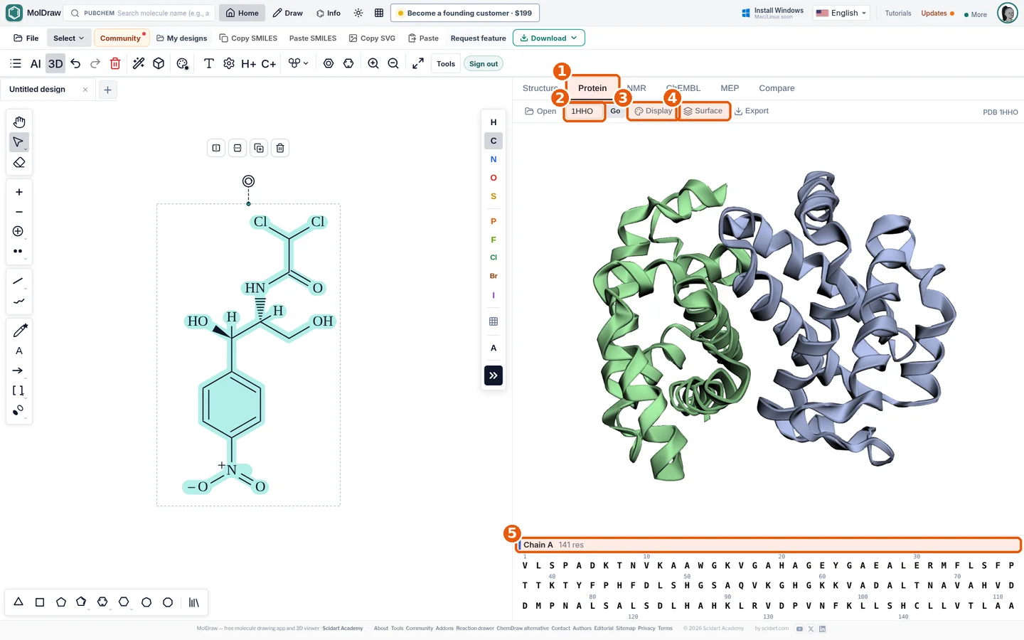
| Load | Type a PDB ID and press Go to fetch it from RCSB, press Open for a local PDB file, or drop a file onto the viewer. |
|---|---|
| Display | Cartoon, Ribbon, Stick, Ball & stick, Sphere, Line or Cross. Colour by Chain, Amino acid, Shapely, Secondary structure, Hydrophobicity, B-factor or Spectrum, or pick your own colour. Set the opacity. |
| Surface | Show or hide a surface, choose SAS, SES, VDW or MS, and set its opacity. |
| Sequence strip | One-letter sequence for each chain. Select residues to give them their own style. |
| Export | GLB, PNG, JPG, PDB, SDF, XYZ, X3D and OBJ, with a Hi-res option. |
How to get the Protein viewer
Need a quick look without opening the editor? The standalone protein structure viewer styles, colours and hides each chain on its own.
You do not need to install anything. The Protein viewer is on by default and works without an account. If you turned it off earlier:
- Open the MolDraw editor.
- Click Settings (the gear), then Plugins.
- Click Install next to Proteins. The Protein tab appears next to Structure.
How to load and style a protein
- Click the Protein tab. A small example structure, crambin (1CRN), loads first.
- Type a PDB ID in the ID box, for example
1HHO, and press Go or Enter. - Open Display. Keep Cartoon and choose Secondary structure to colour helices and strands differently, or choose Chain to tell the subunits apart.
- Open Surface, press Show and lower the opacity so the cartoon stays visible underneath.
- In the sequence strip, click one residue, drag across a range, or Ctrl/Cmd+click to add or remove a residue. In the style panel that opens, set Style to Stick and pick a colour.
- Open Export, tick Hi-res if you need a large image, and choose PNG.
Ways to use it in class and in research
- Teaching protein structure: load 1HHO, colour by secondary structure and point out the helices. Then colour by chain to show that haemoglobin has four subunits.
- Hydrophobic core: colour by Hydrophobicity to show how nonpolar residues tend to sit inside the fold.
- Drug targets: load 6LU7, the SARS-CoV-2 main protease, add a surface and discuss where an inhibitor could bind.
- Figures: style a few key residues as sticks on a cartoon background and export a high-resolution image for a slide or report.
- Next steps: work out a chain's mass with the protein molecular weight calculator, or prepare a receptor for docking with PDB to PDBQT.
Tips and limits
- Good starting IDs: 1HHO (haemoglobin), 1BNA (B-DNA), 6LU7 (SARS-CoV-2 main protease) and 1GFL (green fluorescent protein).
- Each selection you style becomes its own region, so you can colour several sites differently. Use Remove style or Clear selection in the style panel to undo.
- B-factor colouring shows how much atom positions vary in the model. High values often mark flexible loops.
- To open a structure you saved earlier, drag the .pdb file from your computer onto the viewer.
Fetching by ID needs an internet connection, because the file comes from RCSB. The viewer needs WebGL; it shows a message if your browser cannot provide it. The sequence strip lists amino-acid residues only, so ligands, water and nucleic acids do not appear there. A PDB entry is an experimental model, and its quality depends on the method and resolution, which you can check on the entry's RCSB page.
Want to view a small molecule in 3D instead? See how to view molecules in 3D.
Questions and answers
Do I need an account to view proteins?
No. The Protein viewer is installed by default and works without signing in.
Where do the structures come from?
When you type a PDB ID and press Go, the plugin downloads the entry from the RCSB Protein Data Bank. You can also open a PDB file from your computer.
How do I highlight an active site or a few residues?
Select the residues in the sequence strip below the viewer. A small style panel opens where you can switch them to sticks, change their colour or add a surface.
Which export formats are available?
The Export menu offers GLB, PNG, JPG, PDB, SDF, XYZ, X3D and OBJ. Tick Hi-res for a larger image.